Monday, August 26, 2019

Is Exercise really beneficial? Demystifying the myth with Flow Cytometry – CD56+ Natural Killer Cells


Exercise, get moving, get fit, get healthy! Its been a common theme to see posters published by health promotion board everywhere to promote exercising and leading an active lifestyle to prevent cancer. Pfff… Why exercise? Aint no body got time for that. You might change your mind after reading this article.  
Health authorities and practitioners have always vehemently advocated that regular exercise reduces the risk of cancer and metabolic diseases. However, immunological mechanisms linking exercise and reduced cancer risk are limited 
Here in this article, we would look at a small study done in Murdoch University [1], published in 2016 to highlight a few key aspects on how exercise can influence the immune system to potentially contribute to cancer prevention. 

Figure 1: Exercise ellict a bi-phasic response where there is a significant increase in numbers in all lymphocyte groups and a subsequent reduction in the recovery phase.


Simplistically, Figure 1 effectively demonstrated that exercise can trigger a transient bi-phasic response where there is a significant mobilisation of different types of crucial lymphocytes; Natural Killer (NK CD56+) cells, T- (CD3+) and B- (CD19+) lymphocytes into the circulating blood, and then significantly fall in numbers during recovery. This bi-phasic phenomenon has been well documented by multiple studies. Furthermore, as observed in the diagram, the NK cell population is most responsive to an acute bout of exercise. Therefore, this article will focus on the NK cells and how they can potentially contribute to cancer prevention and management.


Figure 2: NK cells are part of the WBC population as seen in the first dot plot diagram, using CD45 as a “mature-cell” marker, we can then select the lymphocyte population as CIRCLED in red. With literature knowledge that NK cells do not express CD14, CD3, CD19, CD20, we can then use the third diagram to NEGATIVELY select the desired population. Subsequently in the final plot we can then further classify the NK cells into their respective subpopulations (CD56dimCD16+ & CD56brightCD16- NK cells)


NK cells are a critical component of the innate arm of the immune system that are able to recognize and eradicate tumor cells without prior antigenic exposure. Phenotypically in flow cytometry terms, they are part of a lymphocyte group that specifically express the CD56 marker and can be classified into CD56dim and CD56bright. In the human body, CD56bright NK cells resides in the secondary lymphoid organs such as lymph nodes and tonsils, whereas the CD56dim NK cells resides in the spleen and blood circulation. Immunologically, the CD56dim population is considered the most mature form of all NK cell variants and has the most tenacious cytotoxicity effect against tumour cells. Moreover, multiple exercise immunology research studies have unanimously demonstrated that the CD56dim NK cells is the most responsive to an acute bout of exercise compared to all other lymphocyte cell types.
An intriguing study done by a group of Danish researchers found that mice who spend more time on a running wheel has their tumors shrunk by 60% compared to their more sedentary mates [2]. Physiologically, a high level of adrenaline and a cytokine known as Interleukin-6 (IL-6) is released into the blood circulation during exercise. Moreover, the researchers also found that adrenaline specifically recruits IL-6 sensitive NK cells and the IL-6 cytokine facilitate attraction of the NK cells to the tumors. Besides that, there is an enhanced expression of NK cell-related activating receptors, stimulatory cytokines and chemokines in the tumors of running mice. Hence the authors suggested that exercise also support the establishment of a tumor microenvironment that is more prone to NK cells activation and consequently promote NK cell cytotoxic destruction of the tumour cells.

This research is hopeful for cancer patients as it provides an inexpensive way to keep their tumor under control. However, some questions remain to be explored, including whether this observation also hold true in humans, or how effective it will be when combined treatment with chemotherapy is carried out in patients.

In conclusion, prevention is better than cure. Exercising is such an economical accessible way to prevent cancer. There is not much negative implications or risks in exercising, but there is a whole world of beneficial health impacts. Most importantly, health is wealth, we should heed the aforementioned advice and take time out regularly to get a bout of moderate intensity exercise. As always, stay fit, stay healthy!
References

  1. Preferential Mobilization and Egress of Type 1 and Type 3 Innate Lymphocytes in Response to Exercise and Hypoxia. Ng Ivan, Fairchild Timothy, Greene Wayne and Hoyne Gerard. Immunome Research 2016.
  2.  Voluntary Running Suppresses Tumor Growth through Epinephrine- and IL-6-Dependent NK Cell Mobilization and Redistribution. Line Pedersen, Manja Idorn, Gitte H. Olofsson, Britt Lauenborg, Intawat Nookaew, Rasmus Hvass Hansen, Helle Hjorth Johannesen, Ju¨ rgen C. Becker, Katrine S. Pedersen, Christine Dethlefsen, Jens Nielsen, Julie Gehl, Bente K. Pedersen, Per thor Straten and Pernille Hojman. Cell Metabolism 2016.
Disclaimer
  • Flow cytometric analysis images depicted in this article are for simplified illustration purposes only and shall not constitute to the exact analysis process.
  • Information provided are for informal reading and not for official research usage

Sunday, July 21, 2019

The B Lymphocytes are ever ready for war! (Part 2)

Human immunity is a complex intricate battalion involving a plethora of different cells cooperating to ensure the health of an individual. Because of the diversity of immune cells, flow cytometry represents the best method for studying functional and phenotypic properties of these cell subsets, especially with the ever-expanding list of Cluster of Differentiation (CD) markers.
The immune system comprises of two distinct arms that are inextricably linked; innate and adaptive immunity. The primary role of the innate immune system is to provide a first line of defence by limiting the colonization of invading pathogens until antigen-specific adaptive immune responses are established.
Since we have previously discussed about the Cell-mediated adaptive T cell immunity (https://www.linkedin.com/post/edit/6479961887206412288), this article will mainly be discussing only on the adaptive humoral immunity B cell and how flow cytometry plays a role. The hallmark of the adaptive humoral immunity is the secretion of powerful multi-functional antibodies that can eradicate bacteria either by neutralisation or promoting opsonisation (phagocytosis/lysis).
The positive selection process of the B lymphoid progenitors occur in the bone marrow where the cells’ surface B Cell Receptor (CD19) is tested for its functionality to complete their maturation as naive B Lymphocytes.
The activation and subsequent maturation of the Naïve B cells is a collaborative effort with the CD3+CD4+ T helper cells. After leaving the bone marrow and upon encountering a deleterious antigen, the Naïve B cell will capture that antigen and present it to a T helper cell via the MHC Class II receptor. This collaboration will elicit an “activation” of the B cell to start differentiating into 2 groups of cells, Plasma B cell or Memory B cell.
The CD138-CD38+ Plasmablast is an immature precursor cell of the Plasma B cell. They have the capability to secrete more antibodies than mature naïve B cells but less than plasma cells. These cells have the flexibility to remain in this state for several days and then either die or differentiate irrevocably into a plasma cell.
The CD138+CD38+ Plasma B cell will start producing antibodies specifically targeting and eliminating the earlier mentioned harmful pathogen. These Plasma B cells are permanently situated at lymphoid organs such as bone marrow, spleen and lymph nodes to produce antibodies to be released into the blood stream.
On the other hand, CD27+IgD- memory B cells are localised in the germinal centers of the lymphatic system. They have one of the longest shelf life in the immunity system like a strong seasoned war-veteran. Their long-lived memory ability to jump-start the immune system to mount a more rapid aggressive response than before against a re-encountered antigen/pathogen is the cornerstone of vaccination. Acting as a recognition center, they will detect any re-encountered pathogen and then phenotypically modulate itself into the aforementioned plasma B cells, to produce immunologic antibodies to eradicate the pathogen.
So here is just a quick snapshot of how flow cytometry aids in studying the phenotypic difference in the various B cell subsets of the humoral adaptive immunity. However, understanding the above phenotypic information is just the tip of an iceberg. Understanding how drugs can affect these lymphocytic population in a diseased state is a long arduous process. Fortunately, the rapid advancement of flow cytometry will be the key to future drug discovery.
Disclaimer
1.      Flow cytometric analysis images depicted in this article are for simplified illustration purposes only and shall not constitute to the exact analysis process
2.      Information provided are for informal reading and not for official research usage

Sunday, July 8, 2018

The Lymphocytes are ever ready for war! (*The Fun Way) - Part 1

Human immunity is a complex intricate battalion involving a plethora of different cells cooperating to ensure the health of an individual. Because of the diversity of immune cells, flow cytometry represents the best method for studying functional and phenotypic properties of these cell subsets, especially with the ever-expanding list of Cluster of Differentiation (CD) markers.
The immune system comprises of two distinct arms that are inextricably linked; innate and adaptive immunity. The primary role of the innate immune system is to provide a first line of defence by limiting the colonization of invading pathogens until antigen-specific adaptive immune responses are established.
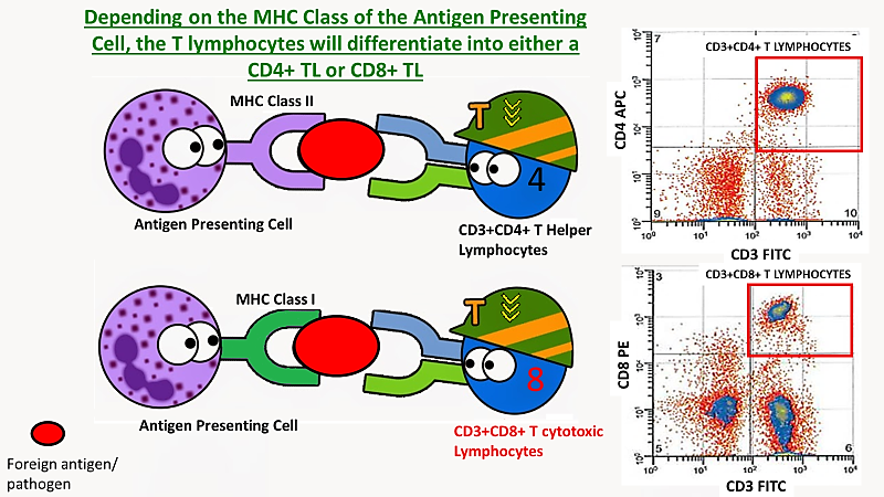
In this article we will be discussing only on adaptive T cell immunity, essentially conferred by the T lymphocytes and how flow cytometry facilitates the in-depth study of lymphocytes. This group of cells have the capability to generate antigen-specific immune responses to a foreign antigen and the ability to induce “immunological memory” that confers rapid and enhanced protection upon subsequent antigen recognition.
The adaptive immune system can be illustrated with an army of soldiers with different “ranks” ready for war. It comprises two classes of specialised cell types which correspond to cell-mediated and humoral immunity respectively: the T-lymphocytes (TLs) and B-Lymphocytes (BLs). T-Lymphocytes that involve cell mediated immunity can be separated into cytotoxic CD8+ TLs and helper CD4+ TLs, which together account for 60-80% of the total lymphocytes in the peripheral immune system. 
 The thymus is a “training facility” where the Lymphoid progenitors undergo a rigorous “selection” process, where their surface T Cell Receptor (CD3) is tested for its functionality. Only cells that express T Cell Receptors (TCRs) with low affinity for self-peptide are positively “selected” to complete their maturation as naive TLs. This process prevents auto-immunity where the T lymphocytes start recognising the host cells wrongly as “foreign” and start attacking them, leading to self-destruction.
So how do the T cell know what is their identity and what type of cell to mature into? Depending on the specificity of the TCR for peptide-MHC Class I or peptide-MHC Class II antigens on antigen presenting cells, this will direct differentiation to either the CD8+ cytotoxic TLs or CD4+ T helper lineage respectively. The role of CD3+CD4+ T helper cells is to secrete cytokines that recruit other immune cells to regulate and enhance immune responses, while CD3+CD8+ Cytotoxic T cells on the other hand produce immunologic chemicals to induce cytotoxicity that aid in the eradication of infected or mutated cells
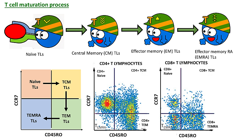
Moving further forward, there are four well characterized major T cell subsets; naïve TLscentral memory (Tcm TLs)Effector memory (Tem TLs) and effector memory RA (Temra TLs). These TLs subsets can be categorized by a number of markers such as CD45RA, CD45RO, CD62L and CCR7. Using 2 surface markers of CD45RO and CCR7 in flow cytometry, we can demonstrate a simplified model to depict the T lymphocyte maturation journey. However, are these surface markers just “facial” features that differentiates one another? Nope! In fact, these surface markers dictate where their position is and where they perform their duties. While some surface markers differentiate who the new recruits are and who the “experienced” soldiers are.
As we all know the more experienced soldiers will get promoted to a higher rank in the army; similarly as the Naïve TLs get exposed to an antigen, they will mature and be promoted to next rank. In flow cytometry terms, the Naïve TLs can be identified by the expression of CD45RA however, once they come into contact with an antigen, they will become “antigen-experienced” and lose the CD45RA expression while gaining CD45RO expression to be “activated” to perform its immunologic function. This is synonymous to dropping the dummy rifle once they passed their training in exchange of a real rifle to begin real war against a real enemy.
As mentioned earlier, immunological memory is the hallmark of adaptive immune system. It enables the immune system to respond more aggressively and rapidly to obnoxious pathogens that have been encountered previously. Two characteristically distinct antigen-experienced CD45RA-CD45RO+ memory TL populations have been widely recognised; the Central memory (Tcm TLs) and Effector memory (Tem TLs). CD62L and CD197 (CCR7) are two common surface markers that distinguishes the two populations.
Expression of CD62L and CCR7 on Central memory Tcm TLs gives them the right to operate and defend within the territory of the lymph nodes. Commonly known as “lymph-node homing markers”, both these markers act similarly like a passport/visa to signify lawful permission to enter or live in a country. Despite lacking immediate effector functions they are able to proliferate and rapidly differentiate into effector cells upon secondary stimulation as well as producing IL-2.
In contrast, the absence of CD62L and CCR7 on Effector memory Tem TLs forbids their entry into the lymph nodes, but express CXCR3 to patrol in the blood stream surrounding the peripheral tissues.
In summary, Tem act as frontiers of immunity against recall Ag, while Tcm is responsible for providing long-term immunity and generating a second batch of effector T cells on secondary challenge.
So here is just a quick snapshot of what the cell-mediated and humoral mediated adaptive immunity is all about. However, understanding the above phenotypic information is just the tip of an iceberg of what human immunity is all about. Understanding how drugs can affect all these lymphocytic population in a diseased state is a long arduous process. Fortunately, the rapid advancement of flow cytometry will be the key to future drug discovery.
Disclaimer
1.      Flow cytometric analysis images depicted in this article are for simplified illustration purposes only and shall not constitute the exact analysis process
2.      Information provided are for informal reading and not for official research usage
3. Views or opinions presented in this post are solely those of the author and do not represent those of the company or manufacturer. The author accepts no liability for the content of this post, or for the consequences of any actions taken on the basis of the information provided.

Wednesday, March 2, 2016

Cell Free DNA – the “floating remnants” that may lead to the hidden wrecks (tumors) under the boundless ocean

Figure 1: Is Cell-Free DNA our next hope to cancer detection?
Do you know that our marine environment is succumbing into severe pollution from corroding shipwrecks? With water making up to 71% of the earth surface, looking for shipwrecks that had sunk into deep waters is synonymous to searching for a needle in a haystack.
Does that actually resemble a kind of disease in our body too? Yes, that is what I am talking about; Cancer and Tumors. While all tumors are not necessarily cancerous, malignant tumors are essentially cancer. Regardless of tumors being benign or malignant, they are basically tissues that are composed of cells whose DNA are mutated, rendering them the abnormal capability to proliferate exponentially without regulation. These usually result in abnormal lesions or swelling of an organ, increasing the competition for “nutrients” with the healthy normal cells, thereby posing potential health hazards to the host. Similarly, removal of these tumor cells will halt the deteriorating health impacts and allow the host to regain its health. Unfortunately, searching for these tumors is not as simple as it seems to be.
It is a widely acknowledged fact that sooner a cancer is diagnosed and treated, better the prognosis. However, a clinically proven circulating biomarker to locate the specific location of the malignant cells remains a mystery. Sure, there are serum-based tumor-markers such as carcinoma antigen-125 (CA-125), carcinoembryonic antigen (CEA), and prostate-specific antigen (PSA) which are traditionally used to detect, diagnose, and manage some types of cancer. Although, the levels of these tumor markers are found to be significantly elevated in cancer patientsit does not confirm cancer diagnosis. Take PSA that is used to screen men for prostate cancer for example.  Though increased PSA level is associated with prostate cancer, there are non-prostate cancer patients with elevated PSA levels.  Therefore measurements of tumor markers are usually combined with other tests, such as biopsies, to diagnose cancer. 
Figure 2: Biopsy needle needed to draw a column of lung tissue to confirm lung cancer diagnosis
Needle-tissue biopsy is traditionally the gold standard for cancer diagnosis. However, this is a highly invasive expensive medical procedure that involves puncturing a needle into the body to obtain tissue samples for more in-depth examination of the cells. Hence, scientists have been diligently researching for the past 2 decades for alternatives that could by-pass needle-tissue biopsy and make cancer diagnosis more efficient and less invasive.
Taking a step back, if floating debris from shipwrecks could lead to the exact location of its host, is there something similar that can aid the physicians to detect the presence of tumors?
Figure 3: Can scientists uncover the mystery of these floating cell free DNA in relevance to Tumors?
Yes, that floating debris in relation with tumors is Cell-Free DNA. In fact, cell-free DNA is not a novel finding. Its existence was first discovered back in 1948 by Mandel and Matis, although debates on the clinical application of cell-free DNA in tumor diagnosis only began in1977.
As the name implies, “cell-free DNA” refers to DNA compounds not found in a cell. Traditionally, DNA is usually localized in protecting organelles such as nucleus and mitochondria. The exciting breakthrough begins when serum DNA levels are significantly elevated in cancer patients compared to healthy individuals, especially in patients with metastatic Tumors. In normal individuals, concentration of cell-free DNA varies from 0 – 100ng/ml of blood.
However, this extravagant surge in cell-free DNA concentration is not limited to cancer patients. Patients languishing in clinical conditions such as rheumatoid disease, trauma, myocardial infarction, fever and inflammatory diseases have similar phenomenon. Although their presence has been well documented in various clinical conditions, the origin and their liberation mechanism remains poorly understood.
Figure 4: Scientists believe that DNA is usually stored safely in the nucleus of cells.
Figure 5: Scientists suspects that when there is a tumor infected tissue adjacent to a blood vessel, there is a tendency for DNA to leak out of the abnormal tumor cells when they undergo necrosis or apoptosis. Their concentration is in relevance to the stage of the cancer.
Majority of the scientists have unanimously believed tumors are a major source of these DNA fragments that are released into the circulation when they undergo necrosis or apoptosis. In addition, a team of scientist observed that the quantity of circulating cell-free DNA correlated with the stage of cancer. With only 47% of the patients in stage 1 cancer having detectable cell-free DNA, the percentage of patients in progressing cancer stages II, III and IV was 55, 69 and 82 percent, respectively. Moreover, they also highlighted that the increasing concentration of cell-free DNA reflects the advancing stages of cancer (1). Therefore, this opens the possibility of determining how advanced a patient’s cancer is just by measuring the circulating level of cell-free DNA.
To further prove the correlation between circulating levels of cell-free DNA and the stages of cancer, Bettegowda and colleagues observed that patients with lower blood levels of cell-free DNA lived significantly longer than those with higher levels of cell-free DNA (2).
Since most studies up to this stage have illustrated that increased cell-free DNA is the hallmark to distinguish cancer patients from healthy individuals, this opens the possibility of using cell-free DNA as an early tumor marker. With the knowledge that tumors arise from genetic mutations, detecting these genetic mutations could be the means to discover an early diagnostic marker. Promisingly, there are a number of studies that have demonstrated tumor-specific genetic and epigenetic changes in the cell-free DNA of cancer patients such as N-ras mutations (acute myelogenous leukaemia (3), K-ras mutations (colorectal carcinoma) (4) and p53 mutations (breast cancer) (5) to name a few.
We now know that concentration of cell-free DNA in the circulation reflects the presence of tumors. The next challenge is to determine the location of the tumors for early cancer detection. There is a definite need to identify a specific sequence or modification on the cell fragments that can specifically identify the source. As a recent breakthrough in 2012, Agostini M and his colleagues found that ALU247can be a specific marker on the cell-free DNA in discriminating breast cancer patients from non-cancer subjects (6).
Since there is so much interest and promising results from the available studies, does that mean, cell free DNA is seeing the light at the end of the tunnel?
Unfortunately, studies regarding the application of cell-free DNA in routine clinical diagnostics are only at its budding stage. In order to succeed, there are some major details to be ironed out. The foremost limitation is that the majority of the research was done with small sample size (n < 100). For clinical application, there is a need to have a huge sample size to smooth out details such as gender differences, racial/national diversities and age group differences. Hence the conclusion derived from the basis of the power of statistical tests used in these studies remain questionable.
Second challenge is the establishment of reference intervals for the healthy population. This aspect requires an immense amount of resources such as extensive monitoring, documentation and meticulous consideration to all variable attributes from pre-analytical to analytical process.
Third issue is the variation observed in the various protocols and assays. Different studies have used different sample collecting tubes, different DNA isolation, quantitation and detection methods, while downstream processing procedures such as centrifugation speed have not been standardized as well too.
Therefore, although cell-free DNA is an exciting breakthrough in the field of clinical diagnostics for early cancer detection and monitoring, there are huge complicated gaps yet to fill. Only when the above limitations are overcome, the “shipwreck – tumors/cancer” will remain secluded, while scientists will continue to “dive” deeper to look for the elusive “debris – cell free DNA”.
Figure 6: Can cell-free DNA be one of the clinical biomarker in our stable of diagnostic assays for early cancer detection and prevention in future? 
References
  1. Spindler KL, Pallisgaard N, Andersen RF, Brandslund I, Jakobsen A. Circulating free DNA as biomarker and source for mutation detection in metastatic colorectal cancer. PloS one. 2015;10(4):e0108247. Epub 2015/04/16.
  2. Bettegowda C, Sausen M, Leary RJ, Kinde I, Wang Y, Agrawal N, et al. Detection of circulating tumor DNA in early- and late-stage human malignancies. Science translational medicine. 2014;6(224):224ra24. Epub 2014/02/21.
  3. Vasioukhin V, Anker P, Maurice P, Lyautey J, Lederrey C, Stroun M. Point mutations of the N-ras gene in the blood plasma DNA of patients with myelodysplastic syndrome or acute myelogenous leukaemia. British journal of haematology. 1994;86(4):774-9. Epub 1994/04/01.
  4. Kopreski MS, Benko FA, Kwee C, Leitzel KE, Eskander E, Lipton A, et al. Detection of mutant K-ras DNA in plasma or serum of patients with colorectal cancer. British journal of cancer. 1997;76(10):1293-9. Epub 1997/01/01.
  5. Silva JM, Dominguez G, Garcia JM, Gonzalez R, Villanueva MJ, Navarro F, et al. Presence of tumor DNA in plasma of breast cancer patients: clinicopathological correlations. Cancer research. 1999;59(13):3251-6. Epub 1999/07/09.
  6. Agostini M, Enzo MV, Bedin C, Belardinelli V, Goldin E, Del Bianco P, et al. Circulating cell-free DNA: a promising marker of regional lymphonode metastasis in breast cancer patients. Cancer biomarkers : section A of Disease markers. 2012;11(2-3):89-98. Epub 2011/01/01.
 Disclaimer: Any views or opinions presented in this post are solely those of the author and do not necessarily represent those of the company or manufacturer. The author accepts no liability for the content of this post, or for the consequences of any actions taken on the basis of the information provided.